
A young man presented with severe pain on the outer side of his knee. Investigations and a biopsy confirmed a giant cell tumor of bone in the proximal tibia, the upper part of the shinbone near the knee. The tumor had weakened the bone and caused a pathological fracture.
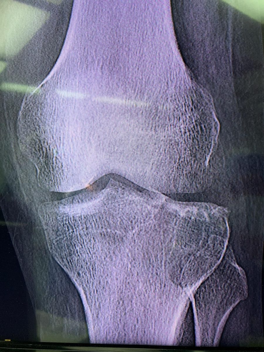
At presentation: the tumor in the upper tibia.
Giant cell tumors are usually benign, but they can behave aggressively where they grow and destroy the surrounding bone. This case shows why a benign tumor can still need complex treatment, particularly when it weakens the bone next to a joint.
Removing the tumor
Our treatment addressed both the tumor and the fracture. We performed curettage to remove the tumor from inside the bone, aiming to preserve the nearby knee joint.
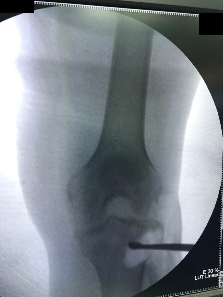
Curettage under X-ray guidance.
A dental burr was used to treat the walls of the cavity, together with argon beam coagulation as a local adjunct. These steps are intended to reduce residual tumor tissue and the risk of recurrence. Argon beam coagulation has been studied as an adjunct to curettage and cementation, although recurrence remains possible.
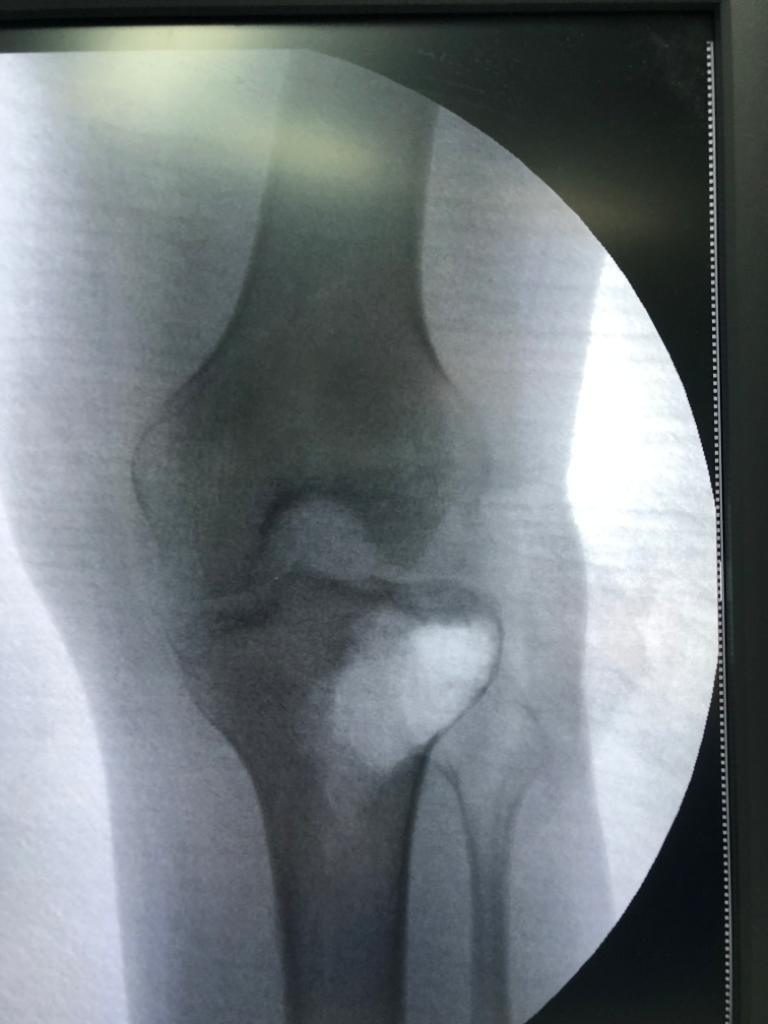
X-ray after the tumor has been fully curetted.
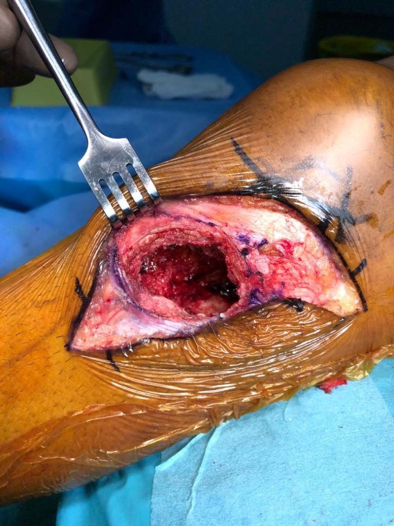
The cavity after curettage, during surgery.
Filling the defect and fixing the bone
After treating the cavity, we filled the defect with bone cement. Cementation provides support within the defect.
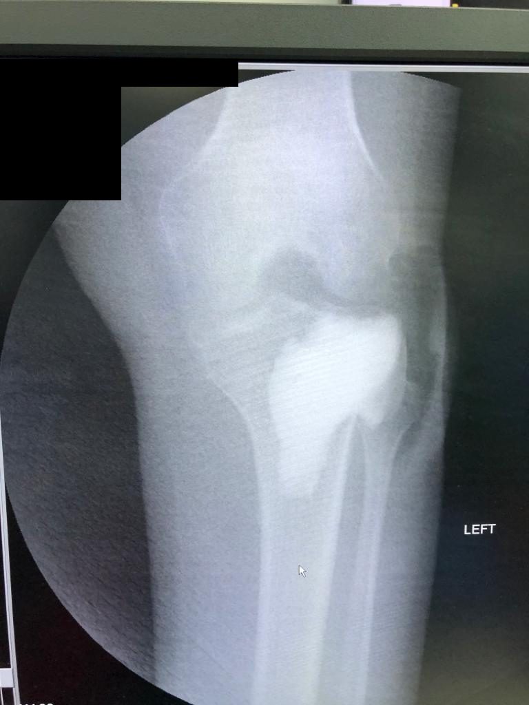
X-ray after filling the cavity with bone cement.
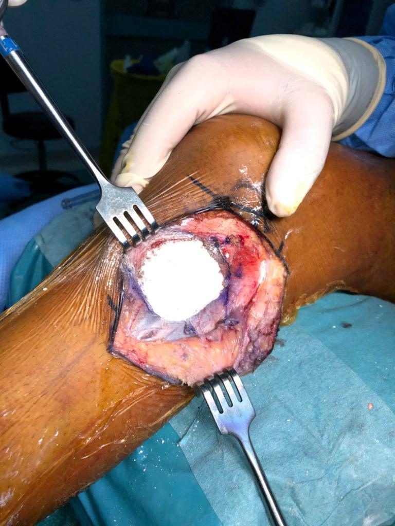
The bone cement in the cavity, during surgery.
The weakened, fractured proximal tibia was then stabilized with an anatomic plate and screws, while the cement supported the defect.
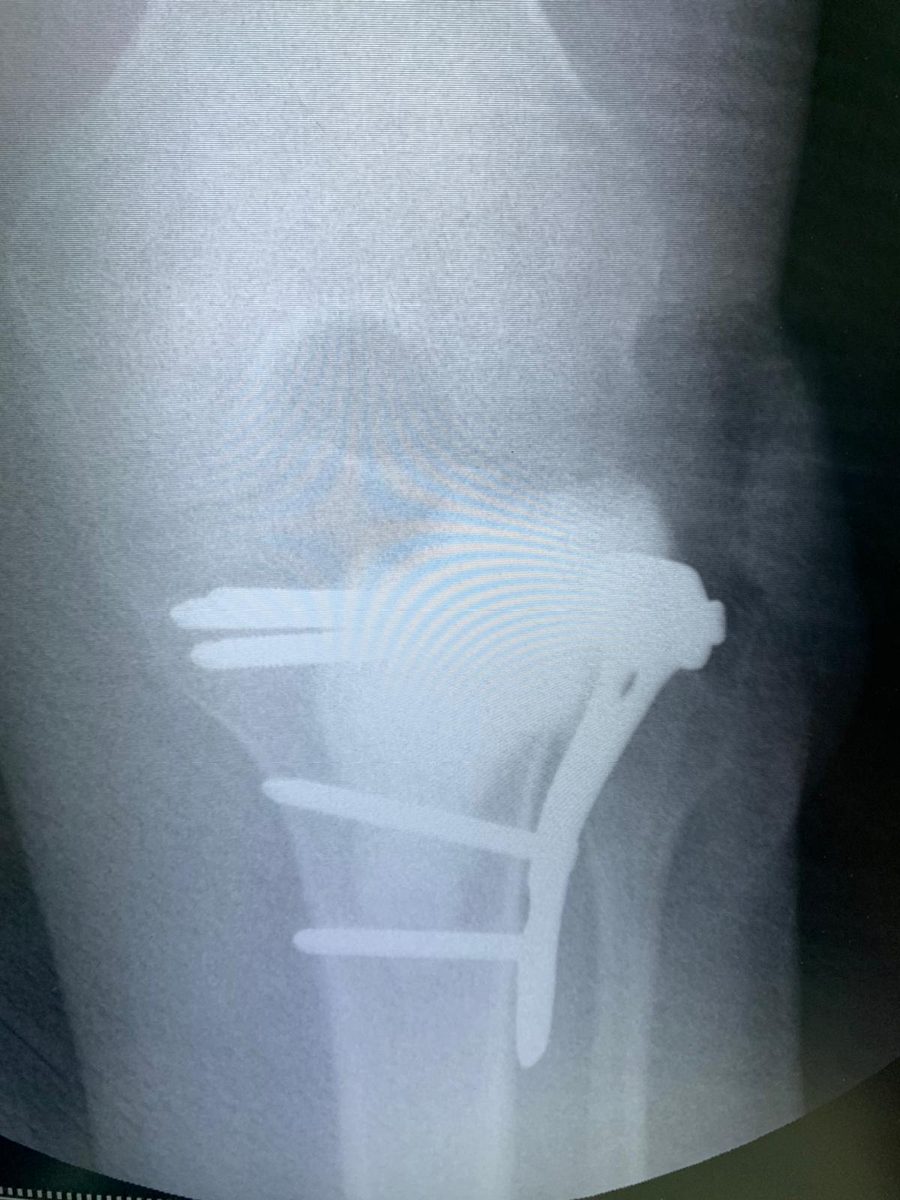
After fixation with the plate and screws.
The aim was to combine local control of the tumor with structural support, while preserving the knee.
Outcome
At three years of follow-up the patient was doing well. He was walking with minimal pain and showed no sign of tumor recurrence. Continued clinical review and imaging remain important, because giant cell tumors can recur after treatment. Rehabilitation and the progression of weight-bearing are individualized.
This case highlights the importance of investigating severe knee pain, confirming the diagnosis with a biopsy, and planning treatment that addresses both the tumor and its effect on the strength of the bone.
Cases are published with the patient's consent and without anything that identifies them. Results differ from patient to patient.
← All cases